Mon - Sat [ 07:00Am - 07:00 PM ] [ Sun: 07:00 AM - 01:00 PM ] 0491 250 1601 / 02 pulsepalakkad@gmail.com
Trusted By
120,000+ People
First Women Clinic
In Palakkad
First Breast Clinic
in Palakkad

DIGITAL MAMMOGRAPHY

BEST MAMMOGRAPHY CENTRE IN PALAKKAD
( The only center in Palakkad town with 3D Mammography with Tomosynthesis )

BEST BREAST SCAN CENTRE IN PALAKKAD. AT PULSE SCANS .
( An initiative of Pulse preventive medicine best practices )
@ PULSE CLINIC .
Home > Our Services > DIGITAL MAMMOGRAPHY

About DIGITAL MAMMOGRAPHY

Mammography is a low-energy X-ray of the breast taken to detect breast disease. A mammogram can detect breast cancer up to two years before it can be felt. Early detection of cancer increases effective treatment options and the possibility of a cure. Mammography has been proven to detect cancer earlier than physical exam alone. .

What if we could find breast cancers earlier?
See lesions more clearly?
Reduce the number of unnecessary biopsies?

We introduce the first 3D mammography system into our little town of Palakkad with the promise of breast tomosynthesis.

Benefits include
  • The fastest breast tomosynthesis scan available- Less than 4 seconds.
  • Less time under compression to improve patient comfort, lowering the risk of motion.
  • Clinically superior exams to conventional 2D imaging.
  • Low radiation dose imaging.
  • 15-degree scan, providing high in-plane resolution.
  • Swift acquisition of projection images enabling rapid reconstruction of high quality images.

Is there any risk in undergoing Mammogram?

The benefits of mammography out weigh any minimal risk .

How Should I Prepare For The Exam?

Do not schedule your Mammogram for the week before your period, as your breasts are usually tender during this time. The best time is one week following your period. Also inform us if you are pregnant. Do not wear deodorant, talcum powder, or lotion under your arms or on your breasts on the day of the exam. These can appear on the X-ray film as calcium spots. If possible, obtain prior mammograms and make them available to the Radiologist. In addition, inform about any prior surgeries, hormone use, family or personal history of breast cancer

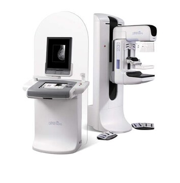
What Does The Mammography Equipment Look Like?

The unit is dedicated equipment because it is used exclusively for X-ray exam of the breast, with special accessories that allow only the breast to be exposed to the X-rays. Attached to the unit is a device that holds and compresses the breast and positions it, so that images can be obtained at different angles.

What is Breast Sonography( Ultrasound)?

Breast Sonography is a painless ultrasound procedure where the breasts are scanned with high frequency sound waves to produce pictures of the breast tissue. In our centre, Mammogram procedure includes a combined breast ultrasound as well, since studies have shown higher accuracy when both are done together and correlated.

Welcome to our breast clinic – best in early breast cancer detection in palakkad Multiple procedure modes offered at PULSE:

  • 2D MAMMOGRAPHY
  • 3D MAMMOGRAPHY
  • Low-dose 3D MAMMOGRAPHY
  • MAMMO GUIDED BIOPSY
  • MAMMO GUIDED WIRE PLACEMENT
  • SONOMAMMOGRAPHY
  • MR MAMMOGRAPHY

At PULSE, we are dedicated to early detection and accurate diagnosis.  Our 3D MAMMOGRAPHY technology offers proven superior clinical performance to 2D mammography alone:


  • Detects 41% more invasive cancers.
  • Reduced false positive recalls by up to 40% .

The Selenia Dimensions system with 3D MAMMOGRAPHY exam provides the groundbreaking technology to allow life-saving treatments to begin earlier.


TOMOSYNTHESIS - A NEW ERA IN BREAST IMAGING


Mammography is specialized medical imaging that uses a low-dose x-ray system to image breasts. A mammogram aids in the early detection and diagnosis of breast diseases in women.

The recent advances in mammography include digital mammography and breast tomosynthesis.

Digital mammography, also called full-field digital mammography (FFDM), is a mammography system in which the x-ray film is replaced by electronics that convert x-rays into mammographic pictures of the breast. These images of the breast are transferred to a computer for review by the radiologist and for long term storage.

Breast tomosynthesis, also called three-dimensional (3-D) mammography and digital breast tomosynthesis (DBT), is an advanced form of breast imaging where multiple images of the breast from different angles are captured and reconstructed( "synthesized") into a three-dimensional image set.

In this way, 3-D breast imaging is similar to computed tomography (CT) imaging in which a series of thin "slices" are assembled together to create a 3-D reconstruction of the body.

The radiation dose for our breast tomosynthesis system remains within the FDA-approved safe levels for radiation from mammograms.

Large population studies have shown that screening with breast tomosynthesis results in improved breast cancer detection rates and fewer "call-backs," instances where women are called back from screening for additional testing because of a potentially abnormal finding.

Breast tomosynthesis may also result in:

  • earlier detection of small breast cancers that may be hidden on a conventional mammogram
  • greater accuracy in pinpointing the size, shape and location of breast abnormalities
  • Faster detection as thin layers of tissue are shown separately and suspicious lesions from a traditional 2D image can be ruled out as benign.
  • Fewer call backs for additional screening and testing;fewer unnecessary biopsies .
  • greater likelihood of detecting multiple breast tumors.
  • clearer images of abnormalities within dense breast tissue.
  • Increased comfort because 2D and 3D images can be produced in a single compression.

Screening Mammography
Mammograms are used as a screening tool to detect early breast cancer in women experiencing no symptoms. Mammography plays a central part in early detection of breast cancers because it can show changes in the breast up to two years before a patient or physician can feel them. Current guidelines from the U.S. Department of Health and Human Services (HHS) and the American College of Radiology (ACR) recommend screening mammography every year for women, beginning at age 40. Research has shown that annual mammograms lead to early detection of breast cancers, when they are most curable and breast-conservation therapies are available.

The National Cancer Institute (NCI) adds that women who have had breast cancer, and those who are at increased risk due to a family history of breast or ovarian cancer, should seek expert medical advice about whether they should begin screening before age 40 and the need for other types of screening.

Diagnostic Mammography
Diagnostic mammography is used to evaluate a patient with abnormal clinical findings—such as a breast lump or nipple discharge—that have been found by the woman or her doctor.

News from the RSNA Journal Radiology

Digital Mammography Superior to Film Mammography
Digital mammography is better screening option than film mammography, according to newly published results from the Digital Mammographic Imaging Screening Trial (DMIST). The study found that digital 3D mammography with tomosynthesis performed better than film mammography for all women and in particular pre- and perimenopausal women under age 50 with dense breasts.

Welcome to our breast clinic – best in early breast cancer detection in Palakkad.