Trusted By
120,000+ People
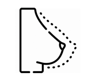

The gold standard for diagnosis of breast disorders
PULSE BREAST CLINIC
provides the region’s
most advanced breast care for women
including routine exams, screening and diagnostic mammograms for the detection of breast cancer, and education about general breast care.
The clinical examination ,breast ultrasound ,mammogram and biopsies are all done in the single visit as far as possible.
Diagnostic facilities offered with state of the art technology and the very best in the world.
We brought in
3D mammography with tomosynthesis
in the year 2017 into Palakkad town ,
the first in a private diagnostic clinic in Kerala.
If you have a family history or personal history of breast cancer or any other conditions that potentially increase your risk for developing breast cancer, contact our clinic for a Risk Assessment.
You may have an increased risk of breast cancer if you have:
Evaluation of breast problems, such as
Best breast scanning centre in Palakkad .
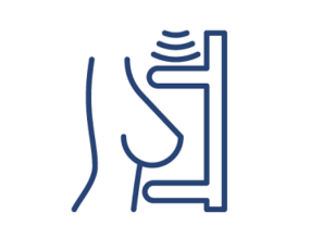
Breast ultrasound uses sound waves and their echoes to make computer pictures of the inside of the breast. It can show certain breast changes, like fluid-filled cysts, that can be harder to see on mammograms.
Ultrasound is not typically used as a routine screening test for breast cancer.
But it can be useful for looking at some breast changes, such as lumps (especially those that can be felt
but not seen on a mammogram). Ultrasound can be especially helpful in women with dense breast tissue,
which can make it hard to see abnormal areas on mammograms. It also can be used to get a better look at a suspicious
area that was seen on a mammogram.
Ultrasound is useful because it can often tell the difference between fluid-filled masses like cysts and solid masses.
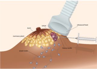
Ultrasound can also be used to help guide a biopsy needle into an area of the breast so that cells can be taken out and tested for cancer.
This can also be done in swollen lymph nodes under the arm.
Ultrasound is widely available and is fairly easy to have done, and it does not expose a person to radiation.
It also tends to cost less than other testing options.
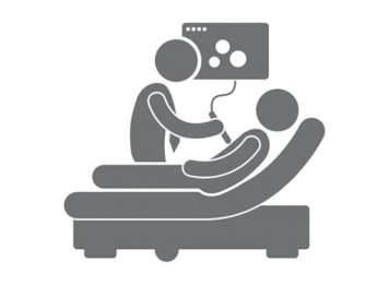
Most often, ultrasound is done using a handheld high frequency transducer. First a gel is put on the skin and/or the transducer, and then the transducer is moved around over the skin. It sends out sound waves and picks up the echoes as they bounce off body tissues deeper under the skin. These echoes are made into a picture on a computer screen. You might feel some pressure as the transducer is moved around on your skin, but it should not be painful.
Doctors use the same standard system to describe results of mammograms, breast ultrasound, and breast MRI.
This system (called the Breast Imaging Reporting and Data System or BI-RADS) sorts the results into categories
numbered 0 through 6.
By sorting the results into these categories, doctors can describe what they find on an ultrasound using the same
words and terms. This makes communicating about these test results and following up after the tests much easier.
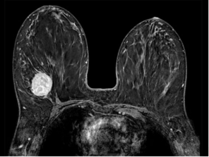
Best breast MRI centre in Palakkad
Breast MRI ( magnetic resonance imaging ) uses radio waves and strong magnets to make detailed pictures of the inside of the breast.
Breast MRI might be used in different situations.
To screen for breast cancer :
For certain women at high risk for breast cancer, a screening breast MRI is recommended along
with a yearly mammogram. MRI is not recommended as a screening test by itself, because it can miss
some cancers that a mammogram would find.
Although MRI can find some cancers not seen on a mammogram, it’s also more likely to find things
that turn out not to be cancer (called a false positive). This can result in some women getting tests
and/or biopsies that end up not being needed. This is why MRI is not recommended as a screening test
for women at average risk of breast cancer.
To look at the breasts if someone has symptoms that might be from breast cancer :
Breast MRI might sometimes be done if breast cancer is suspected (based on symptoms or exam findings,
such as suspicious nipple discharge). Other imaging tests such as mammograms and breast ultrasound are usually
done first, but MRI might be done if the results of these tests aren’t clear.
To help determine the extent of breast cancer :
If breast cancer has already been diagnosed, breast MRI is sometimes done to help determine the exact size and
location of the cancer, to look for other tumors in the breast, and to check for tumors in the other breast.
Breast MRI isn’t always helpful in this setting, so not every woman who has been diagnosed with breast cancer needs this test.
To check for silicone breast implant leaks :
In women with silicone breast implants, breast MRI can be used to check for implant leaks.
This isn’t used for women with saline breast implants.
Just as mammograms are done using x-ray machines specially designed for the breasts,
breast MRI also requires special equipment. This MRI machine has a special device called a
dedicated breast coil to image the breasts.
MRI uses strong magnets instead of radiation to make very detailed, cross-sectional pictures of
the body. An MRI scanner takes pictures from many angles, as if someone were looking at a slice
of your body from the front, from the side, or from above your head. MRI creates pictures of soft
tissue parts of the body that would sometimes be hard to see using other imaging tests.
Unlike mammograms or breast ultrasound, breast MRI requires that you have a contrast dye injected
into your vein (through an IV line) before the pictures are taken. This helps make any abnormal areas
in your breasts easier to see.
Follow all instructions:
You don’t usually need a special diet or preparation before an MRI.
Fix an appointment and follow any instructions you’re given.
If you have trouble with enclosed spaces:
Breast MRI is most often done while you are lying on your belly with your arms above your head inside a long,
narrow tube. If being in a tight space might be a problem for you, you might need to take medicine to help you
relax while in the scanner. Talking with the technologist or a patient counselor or getting a tour of the MRI machine
before the test can also help. You’ll be in the exam room alone during the test, but you can talk to the MR technologist,
who can see and hear what’s going on.
Remove metal objects:
Before the test, you'll be asked to undress and put on a gown or other clothes without zippers or metal. Be sure to
remove any metal objects you can, like hair clips, jewelry, dental work, and body piercings.
Let your technologist know if you have any medical implants or clips in your body. If you have any of
these types of medical implants, you should not even enter the MRI scanning area unless you're told it's OK to do
so by a radiologist or technologist:
MRI scans are usually done in an outpatient setting in a hospital or clinic. You'll first have an IV line
placed a vein in your arm so that contrast material can be injected during the test.
You’ll lie face down on a narrow, flat table with your arms above your head. Your breasts will hang
down into an opening in the table so they can be scanned without being compressed. The technologist may use
pillows to make you comfortable and help keep you from moving. The table then slides into a long, narrow tube.
When breast MRI is done to look for breast cancer, a contrast material called gadolinium is injected
into a vein in the arm during the exam, which helps show any abnormal areas of breast tissue.
(This is different from the contrast dye used in CT scans.) Let the technologist know if you have any allergies
or have had problems before with any contrast or dye used in imaging tests.
The test is painless, but you have to lie still inside the narrow tube. You may be asked to hold your breath
or keep very still during certain parts of the test. The machine may make loud thumping, clicking, and whirring noises,
much like the sound of a washing machine, as the magnet switches on and off. Some facilities give you earplugs or headphones
to help block noise out during testing.
It’s important to stay very still while the test is being done, which helps ensure the images will be of good quality.
Each set of images usually takes a few minutes, and the whole test usually takes about 30 to 45 minutes. After the test,
you may be asked to wait while the pictures are checked to see if more are needed.
For a newer MRI technique, known as abbreviated breast MRI, fewer images are taken, so the scan takes less time (usually
about 10 minutes).
Doctors use the same standard system to describe results of mammograms, breast ultrasound, and breast MRI. This system (called the Breast Imaging Reporting and Data System or BI-RADS) sorts the results into categories numbered 0 through 6.
Best breast centre in Palakkad for guided biopsy / FNAC .
Breast Biopsy
If breast symptoms or the results of an imaging test (such as a mammogram) suggest you might
have breast cancer, you may need a breast biopsy.
A biopsy of the breast is a procedure done to remove a piece of breast tissue or tumor,
called a sample. During a biopsy, a doctor takes samples from the suspicious area so they
can be looked at in the lab to see if they contain cancer cells.
Needing a breast biopsy doesn’t necessarily mean you have cancer. Most biopsy results
are not cancer, but a biopsy is the only way to find out for sure.
There are different kinds of breast biopsies. Some are done using a hollow needle, and some use an incision (cut in the skin). The type you have depends on a number of things, like:
Screening refers to tests and exams used to find a disease in people who don’t have any symptoms. The goal of screening
tests for breast cancer is to find it early, before it causes symptoms (like a lump in the breast that can be felt).
Early detection means finding and diagnosing a disease earlier than if you’d waited for symptoms to start.
Breast cancers found during screening exams are more likely to be smaller and less likely to have spread
outside the breast. The size of a breast cancer and how far it has spread are some of the most important factors
in predicting the prognosis (outlook) of a woman with this disease.
American Cancer Society screening recommendations for women at average breast cancer risk
These guidelines are for women at average risk for breast cancer. For screening purposes, a woman is considered
to be at average risk if she doesn’t have a personal history of breast cancer, a strong family history of breast
cancer, or a genetic mutation known to increase risk of breast cancer (such as in a BRCA gene), and has not had
chest radiation therapy before the age of 30. (See below for guidelines for women at high risk.)
Women between 40 and 44
have the option to start screening with a mammogram every year.
Women 45 to 54
should get mammograms every year.
Women 55 and older
can switch to a mammogram every other year, or they can choose to continue yearly mammograms.
Screening should continue as long as a woman is in good health and is expected to live at least 10 more years.
All women should understand what to expect when getting a mammogram for breast cancer screening – what the test can and cannot do.
( COURTESY : AMERICAN CANCER SOCIETY FOR USG ; / MRI MAMMOGRAM ; GUIDED FNAC / BIOPSY AND SCREENING RECOMMENDATIONS . )
PULSE BREAST PACKAGES ABOVE 35 YEARS
PULSE BREAST PACKAGES BELOW 35 YEARS