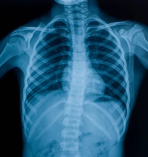
An X-ray image is created when a small amount of radiation is passed through a body part.
Different internal structures absorb different amounts of radiation, which is why the end result of an
X-ray shows solid bone structures and metals as white ; air in lungs as black and fat and muscle
as shades of grey .
X-ray is the most common form of medical imaging.
An X-ray is a quick, painless test .
A very common imaging test done for various indications.
Few of the indications are
Some people worry that X-rays aren't safe because radiation exposure is harmful and may lead to
cancer. The amount of radiation you're exposed to during an X-ray depends on the tissue or organ
being examined. Sensitivity to the radiation depends on your age, with children being more
sensitive than adults.
Generally, however, radiation exposure from an X-ray is low, and your physician while requesting
for the test is aware that the benefits from these tests far outweigh the risks .
Specifically, we use low radiation protocols routinely.
However, if you are pregnant or suspect that you may be pregnant, tell your doctor before
having an X-ray.
Most routine x rays require no preparation ; however a few special types of X-rays / procedures
require different preparations.
Ask your doctor or nurse to provide you with specific instructions.
Feel free to contact us for the preparation required.
Taking appointments is the most desirable ; however we should be able to fit you in the day.
Appointments compulsory for contrast procedures.
In general, you undress whatever part of your body needs examination. You may be given a gown during the exam, depending on which area is being X-rayed. You may also be asked to remove jewelry, eyeglasses and any metal objects because they can show up on an X-ray.
During the X-ray
If a young child is having an X-ray, restraints or other techniques may be used to keep him or her
still. These won't harm your child and will prevent the need for a repeat procedure, which may be
necessary if the child moves during the X-ray exposure.
You may be allowed to remain with your child during the test. If you remain in the room during the
X-ray exposure, you'll likely be asked to wear a lead apron to shield you from unnecessary
exposure.
X-rays are saved digitally on computers, which can be viewed on-screen within minutes.
We have specialist radiologist to view and interpret the results .
Your DIGITAL XRAY results can be made available to your doctor in minutes usually.
After an X-ray, you generally can resume normal activities.
Routine X-rays usually have no side effects.
You will be given an image of the X – ray in approximately 15-20 minutes.
A report will be issued on request ,usually within two hours.
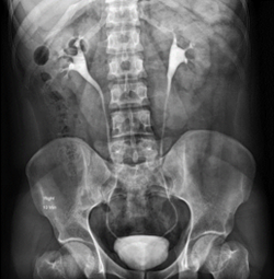
An intravenous pyelogram also called an excretory urogram, is an X-ray exam of your urinary tract. An intravenous pyelogram delineates your kidneys, your bladder and the tubes that carry urine from your kidneys to your bladder (ureters).
During an intravenous pyelogram, you'll have an X-ray dye (iodine contrast solution) injected into a vein in your arm. The dye flows into your kidneys, ureters and bladder, outlining each of these structures. X-ray pictures are taken at specific times during the exam, so your doctor can clearly see your urinary tract and assess how well it's working.
An intravenous pyelogram is used to examine your kidneys, ureters and bladder. In the past, intravenous pyelogram was the most frequently used imaging test for evaluating possible urinary tract disorders. Since the development of kidney (renal) ultrasound and CT scans — which take less time and don't require X-ray dye — use of intravenous pyelograms has become less common.
However, an intravenous pyelogram still can be a helpful diagnostic tool, particularly for:
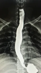
A barium X-ray is a radiographic (X-ray) examination of the gastrointestinal (GI) tract. Barium X-rays (also called upper and lower GI series) are used to diagnose abnormalities of the GI tract, such as tumors, ulcers and other inflammatory conditions, polyps, hernias, and strictures.since the development of ultrasound ,CTand MRI scans ; barium studies have become less common.
Barium is a dry, white, chalky powder that is mixed with water to make barium liquid. Barium is an X-ray absorber and appears white on X-ray film. When instilled into the GI tract, barium coats the inside wall of the esophagus, stomach, large intestine, and/or small intestine so that the inside wall lining, size, shape, contour, and patency are visible on X-ray. This process shows differences that might not be seen on standard X-rays. Barium is used only for diagnostic studies of the GI tract.
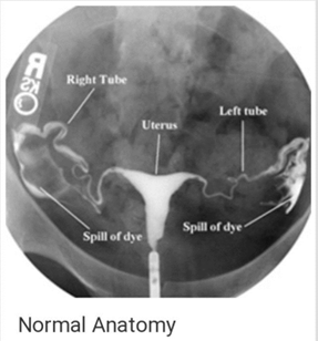
Hysterosalpingography is an x-ray examination of a woman's uterus and fallopian tubes that uses a contrast material.
HSG is not done if a woman has any of the following conditions:
A woman is usually referred for an HSG by her fertility team.
Discomfort – patients’ pain thresholds vary. Some women don’t feel a thing, some find it uncomfortable a bit like period cramps. Only a very few, in our experience, find it very painful.
Infection – this is uncommon and most likely to occur in women who have had an infection before .
After HSG, you can expect to have a sticky vaginal discharge as some of the fluid drains out of the uterus. The fluid may be tinged with blood. A pad can be used for the vaginal discharge. Do not use a tampon.
You also may feel dizzy / faint / or sick to your stomach.
The patient may experience vaginal spotting (bleeding) and pelvic cramps for 1 day after the procedure.
Analgesics and antibiotics will be prescribed as necessary.
Severe problems after an HSG are rare.
They include an allergic reaction to the dye, injury to the uterus, or pelvic infection. Contact your doctor if you have any of these symptoms:
There have been many anecdotal reports of enhanced natural pregnancy rates after an HSG.
So, in couples with unexplained infertility the HSG is not only diagnostic but may also offer them an increase chance of natural conception.
For some types of X-ray tests, a contrast medium — such as iodine or barium — is introduced into your body to provide greater detail on the images.
Before some types of X-rays, you're given a liquid called contrast medium. Contrast mediums, such as barium and iodine, help outline a specific area of your body on the X-ray image. You may swallow the contrast medium or receive it as an injection or an enema.
Please bring a list of your current medications with you (out-patient).
Do inform technician of any allergy to food / medicine.
In some people, the injection of a contrast medium can cause side effects such as:
If you notice above symptoms ,please contact your doctor immediately.
However, if you're injected with contrast medium before your X-rays, drink plenty of fluids to help rid your body of it.
Call your doctor if you have pain, swelling or redness at the injection site.